The Anatomy of Movement – The Vertebral Column
by Dr. Quentin N. LaHam
Taken from the archives of The Canine Chronicle
For several years I taught Comparative Anatomy to biologists and in that capacity I had the occasion to dissect many different animals and to assist hundreds of students wrestling with the problem for the first time. The dogs employed were obtained from the local humane society following euthanasia. They were of medium size for the most part and of mixed breed origin.
The striking feature of this diversity was that despite the variation in the conformity of the individuals the general body plan remained consistent. For example, the top of the shoulder blades were always placed between the first and fifth Thoracic (dorsal) Vertebrae and this was not influenced by the degree of angulation of the individual. This and other structural features led me to conclude that an individual’s vertebral column greatly influences the animal’s final structure and that many of the virtues or shortcomings observed in our dogs can be directly or indirectly related to it.
Later I learned that I was not the only one to hold this view when I chanced across a book The Anatomy of Dog Breeding by Smythe, a British veterinarian, who stated: “If man ever succeeds in producing an animal possessing a perfect spine he will have gone very far toward producing the perfect animal.”
I wish to underscore this statement because too often I have found breeders going after the solution to a problem from the wrong direction. For example, is the apparent short neck on a dog really a short neck or is the problem the position of the shoulders? As shall be seen in subsequent articles, if that is the case it is likely the thorax is the culprit whereas the neck is correct.
It is my strong held view that it is imperative that we learn as much as possible about the vertebral column because in so doing we will learn much about the structure of the dog.
The diversity among mammals is obvious to everyone. This vast array of animals, all of whom suckle their young is common knowledge to most of us from early childhood.
While it may seem strange that animals with such totally different appearances belong to the same class, it should come as no surprise that their vertebral columns have the same primary divisions even though there are differences in the number of individual vertebrae in all divisions except the neck.
The Divisions of the Vertebral Column
There are five regions to the vertebral column. They are:
- The Cervical (Neck)
- The Thoracic (Thorax)
- The Lumbar (Loin)
- The Sacral (Pelvic)
- The Coccygeal (Tail)
Fortunately, all dogs regardless of size or shape normally have the same number of vertebrae in each region except for the tail where the number and shape represents the greatest deviation from the general plan.
Since I consider knowledge of the vertebral column to be so important, this and succeeding articles will be devoted to a single region.
The Neck of the Dog
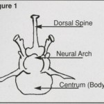
Interestingly, the number of neck vertebrae is consistent in all mammals. Whether it be the long-necked giraffe, the seemingly neckless whale, the dog, the cat or ourselves – we all have seven cervical vertebrae. In anatomy, they are referred to as C1-C7. As each area has differences in the individual vertebrae of the chain associated with their function, the neck is designed to carry, support and move the head at the end of this cantilevered structure. Its vertebrae differ in many ways from those behind and even from each other. To assist in clarifying look at figure 1 which is a simplified schematic presentation of a typical vertebrae
The dorsal spine projects upward from the fusion of the neural arches which in turn encase and protect the spinal cord passing through it. The transverse processes extend outward from the junction of the neural arches with the body of the vertebrae and serve as attachments for muscles and ribs. The body is a bony structure constituting the main mass of the vertebra which together with the intervertebral discs align the vertebrae binding them together in the chain.
There is more to a vertebrae than this simple drawing illustrates which will become clear when we examine them more closely.
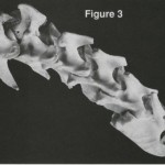
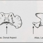
As you read on make repeated referrals to the top (dorsal) view and the side (lateral) view of figures 2 & 3 to help you appreciate the uniqueness of the design of the neck vertebrae and how much they differ from the “typical” design.
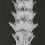 Notice that C1 and C2 not only differ from each other but in turn are very different from the remaining five which more closely resemble each other.
As a group we can make the following observations:
They are broad on their dorsal and ventral surfaces but are relatively narrow from top to bottom; individually, with the exception of C1 they are long and the transverse processes are short, expanded at their ends and very strong. Not shown because they were destroyed in preparing the skeleton are ligaments that help to bind the vertebrae together throughout the length of the column.
Why such massive vertebrae with their large surface area in the neck?
The answer is readily understood and we need only a limited knowledge of the relationship of muscle to bone and some simplified knowledge of muscle anatomy and physiology to comprehend it.
Commit the following to memory as it will serve you well from this point throughout many of the chapters to follow.
- You can only have as much muscle as you have surface area upon which to attach it.
- A muscle has three gross anatomical parts:
a) The “origin” which is the end of the muscle whose tendon is attached to the point which does not move when the muscle contracts.
b) The “insertion,” the opposite end of the muscle whose tendon attaches to a point which moves when the muscle contracts.
c) The “body or belly” of the muscle which is the main mass that contracts or extends dependent upon the action desired. Keep in mind that the muscle that brings about skeletal movement is “voluntary muscle”, that is, it is under control of the individual whether or not it is to contract.
- A muscle can maximally contract (shorten) approximately 50%. In other words, in theory a muscle twelve inches long when maximally contracted would shorten to six inches resulting in pulling the insertion toward the origin to its full extent.
- Very few actions of voluntary muscle involve the action of a single muscle: some eye muscles fall in this category. Most actions are brought about by the combined action of two or more muscles in which case the muscles are said to act synergistically. Synergisms are common in nature but if remembering this word is difficult I’ve always thought if you must sin its usually more pleasurable with somebody else!
- For every action of a muscle or group of muscles there is one or more to do exactly the opposite. Commonly stated for every action there is an equal and opposite reaction.
- When one group of muscles does the opposite to another they are said to be antagonistic to one another.
An illustration should make all the above clear. When you “bend your elbow” two muscles, the biceps (two heads) and the Brachialis muscle are involved (synergism). The origin of these two muscles is on the upper end of the upper arm, (humerus); the insertion is on the middle of the lower arm (radius and ulna). When you attempt to maximally contract these muscles to lower the arm and hand are brought (flexed) against the upper arm. In so doing the muscles have shortened causing our biceps to noticeably bulge. Meanwhile, the triceps (three-headed muscle) has passively elongated to allow this to occur because its role is antagonistic to the others since when it contracts we straighten out our lower arm and again the origin is on the humerus and the insertion on the radius and ulna, but on the opposite side.
Back to the neck. Given the description of the neck vertebrae it becomes obvious that length of neck is determined by the length of the seven vertebrae and the intervertebral discs (cushions) between them. Bear in mind that these vertebrae are determined by a specific, complex set of genes that determines not only their length but the individual differences between this chain of bones, all of which must be taken into account when selecting breeding partners.
Notice also that the individual vertebrae articulate with each other by joints located toward the outside. Having an articulation on either side, rather than a single central articulation, gives much more lateral stability to the neck. Also, note that there is very little in the way of a dorsal spine until the seventh. The absence of dorsal spines allows for greater flexibility of the neck so that the dog can more easily make the adjustments necessary when raising and lowering the head or arching the neck. Remember that the neck vertebrae have a large surface area which is very important because it is directly related to the amount of muscle that can develop in the area. Since the head is comparatively heavy with its weight at the extreme end of the cantilever, strong muscles are required just to hold it up. We know this to be true from our own experience. When we’re tired, we find it difficult to hold up our head. There are, however, other reasons as we shall see shortly for having large, broad neck vertebrae which concern controlling the center of gravity and extension of the forearm.
The first neck vertebrae (C1) is called the Atlas from the greek god because it must be strong enough to support the globe (head). Basically, it is a bony ring with wing-like structures on either side which provide a large area for muscle attachment. It’s articulation with the skull allows for extension and flexion of the head for which it is often called the “yes joint”. It’s articulation with the second vertebrae permits rotary movement and is called the “no joint”. The second vertebrae (C2) shows the axis is by far the most massive. It reminds me of the chest and back plate of an armored medieval knight. In addition to its size it has other unique features. On its anterior ventral surface, there is a finger-like projection that lies in a groove inside the bottom of the ring of the atlas. (figure 4) In human anatomy this is sometimes referred to as “hangman’s notch” because at the time of hanging, proper placement of the noose ensured that the snap of the neck will drive this process into the spinal cord resulting in paralysis and quicker death. A tumbling dog can suffer a similar fate.
The point has been repeatedly made that the larger the cross-sectional area of bone the greater the mass of muscle that can develop and attach thereto. Whenever strength is of paramount importance, we find heavier, larger bones; when speed and agility are paramount, strength is sacrificed and bones tend to elongate and narrow. Thus, the difference between a work horse and a race horse or the bulldog and the whippet. Depending upon what we wish to achieve in a given breed, we make tradeoffs accordingly.
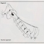
On the dorsal side (top) of the vertebrae on either side of center run bands of worm-like muscles. They run from one end of the vertebral column to the other attaching the different vertebrae and the rib along the way. These groups have different names in the various regions but for our purpose they can be thought of as a band of muscle on either side of the vertebrae. Their function is to strengthen the vertebral column along its axis. The presence of broad vertebrae in the neck make it possible to have strong supportive muscles for the long lever with the head at the end. In addition to the muscles and small ligaments that help to bind the vertebrae together, an elastic cord runs from the dorsal spines of the withers’ region to the axis vertebrae. (figure 5) This interesting tissue being elastic is capable of stretching when the head moves downward and returning to its original shorter length when the tension is released. This elasticity obviously helps the dog to support and raise its head. It is interesting to note that the size of this ligament is structurally related. Thus, bovines and horses have very well-developed elastic ligaments to accommodate their larger, heavier heads. In our dogs, the heavier headed breeds have thicker, broader ligaments.
Our breed standards, when talking about the neck, refer almost universally to the arch of the neck which then slopes gradually to the shoulders. The arch is the result of the larger mass of muscle that attaches to the neck vertebrae in particular to the atlas and axis. There is no way that a dog can have a nice arch to the neck unless the atlas and axis are well developed and, if they are, so too will be the remaining five.
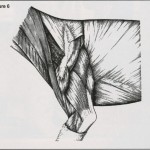
That explains how we achieve that lovely arch but it does not explain why we want it so well-developed. The muscles responsible for the arch are mainly responsible for the extension of the front legs. There are two strap–like muscles (Brachiocaephalicus & Omotransversarius) that run from the base of the skull, the atlas and axis vertebrae, along the dorso-lateral side of the neck to attach to the anterior surface of the upper arm and point of the shoulder respectively (figure 6). To better understand their role in movement, recall the six points I made about muscles. Especially recall that the length of the muscle can shorten maximally by one-half. Comparing a twelve-inch long muscle to one fourteen inches means a difference of one inch of shortening. This may seem not to be a “big deal” but translated to its effect on the inserted lever it becomes an order of magnitude in terms of reach. Thus, the desirability of a longer rather than shorter neck in order to have greater reach when in combination with corresponding length of leg.
 To give you some feeling for the role of the two strap muscles referred to above, try the following experiment upon yourself. First, however, you should be aware that what you are about to do will give you the overall feeling for what these muscles can do but it will not be identical to the action of a dog because you have a collar bone (clavicle). In jumping animals, the clavicle is reduced or absent. In the average-sized dog, it is a small, thin fibrocartilage structure ¾”-1-inch long, about the width of the blunt end of a toothpick and it is embedded in the Brachiocaphalic (arm to head) strap muscles. What this experiment will do is allow you to feel the pull of these muscles in your neck. Perform the experiment slowly and gradually as you are only to feel the pull of the muscle not to inflict pain and cause a stiff neck!
Sit upright in a straight chair, then bend your body to one side, dropping your shoulder bend your elbow and take hold of the chair so it is not possible to straighten up. Now slowly bend your head toward the opposite side as though to place it upon that shoulder. Do not hurt yourself! You should have felt the pull or a twinge if you did it properly.
When your dog elevates and flexes his shoulder and elbow to get his paw off the ground, he now has to extend it forward. This is accomplished by involving several muscles but principally the two strap muscles and the superficial pectoral muscles. The latter run from the two segments of the breast bone from the first two segments (sternum) to the inner side of the upper arm. Pectoral refers to the forelimb, for the same reason we refer to the pectoral fins of the fish. Their role is to hold the forelimb close to the body (adduction) while it is extended (protracted). The principle extensors of the forearm, however, are the two-strap muscle and the dog controls their tension, length and direction of pull by the positioning of his head and extension of his neck. Although the dog is on all fours, we can draw the analogy between a person running and the dog gaiting. When we stand at ease or the dog is in normal stance, both are in as close as possible to a state of static balance, i.e., the body weight is distributed so as to position the center of gravity as close as possible under the weight bearing legs.
 What happens when we walk or even when we jog? We tend to keep our back, shoulders and head erect in order to keep the center of gravity in as straight a line as possible through our body. The sprinters or hurdlers cannot do that if they hope to win. At that moment, in order to increase speed they change from a condition of kinetic balance to that of kinetic imbalance. They lean so far forward that if they didn’t pick up speed and extend their legs they would fall on their faces! So too our dogs, if out for a stroll or a reconnaissance trot, the dog carries his head higher and usually surveys his surroundings. What does he do when he sees something he wishes to investigate? He progressively lowers his head and stretches his neck to put himself in a state if kinetic imbalance until when he’s at a flat out trot his head is lowered to approximately the level of his topline and his neck is fully extended and so too the front legs. One can often observe this in the show ring when in a class of dogs one behind takes a keen interest in the dog in front and reaches out as though he’d like nothing better than a mouthful of his tail!
We have a serious problem in the show ring today because too many dogs are restrained by their handlers who use the leash as though it were a bearing rein. They call upon the dog to increase his speed while forcing his head up. Little wonder the front feet lift too far off the ground in a faschist salute, a hackney, or a windmill motion. Then there are those infamous handlers of German Shepherds, and more recently other breeds, as well who think the name of the game is to see which one can run the fastest. Perhaps leash makers should hire certain German Shepherd handlers to test their load capacity because the dogs are trained to drag their handlers in the ring as though trying to see if they can possibly break the snaps. It is inexcusable to properly assess the dog’s movement, especially coming and going. It might be a good idea for judges to excuse these dogs with the notation “unable to examine”. Unfortunately to do so would undoubtedly require, at best, a flurry of paperwork from the governing bodies or/and bench show hearings and support from other handlers who would claim it wasn’t true!
Another problem confronted in the ring which adversely influences proper use of the head and neck in movement is what I refer to as the “Liver Syndrome.” At the rate it is used in the ring, I wouldn’t be surprised to find pigs on the endangered species list. No doubt you have observed the direct relationship between the size of dog and the handler’s liver pouch. We have small ones for toy breeds, progressively large ones as the size increases until in the larger breeds saddlebags are the in thing!
Somewhere over the years the purpose of baiting a dog has been lost. There was a time when a piece of liver would last a handler all day with some left over. It seems to me that nowadays showtime has become feeding time. The common result is that some dogs run around the ring without the slightest concern for where they are going. They have a one-track mind which repeats over and over, “Where’s the liver, where’s the liver?” He is so interested that he twists his body to crab or sidewind completely distorting his movement.
There are many things the judge looks for when the dog is coming or going and one of them is to see if he can move in a straight line without leash restraint as this is one of the better ways to determine if the front and rear of the dog are in balance. It is very frustrating for the judge to see what is thought to be a good and well-balanced dog distorted by bad handling practices. I’m sure all the handlers of low-stationed dogs would like to “kill” for what happens to their dogs if the breeds preceding them litter the ring with their particular baiting tidbits. They become the self-appointed cleanup crew as every few steps their dogs lunge for a “tasty” morsel.
I am not trying to be flippant; I merely wish to bring home the point that, when used properly and at the proper time, baiting can bring the dog to his best presence but its abuse causes or exaggerates movement faults because of what they do to the head and neck and the muscles that control it. Think of how hard it must be for the dog to run straight forward with the head and neck turned to the right with additional weight shifted to the left front leg.
 To give final emphasis to the role of the two strap muscles, give some thought to this fact. We talked of muscles having an origin and an insertion but the fact of the matter is that in the case of these two muscles (the point that does not move) is the base of the skull and the atlas and axis; the point of insertion is the front of the upper arm and shoulder because the arm is drawn toward the head. What happens when the dog wants to get a flea or go after an irritation on his backside or tail? What he does is pull his front leg back (we’ll see how later) and places it firmly on the ground. At this point the origin of the two strap muscles shift to the upper arm and their insertion to the head and neck. Now when these muscles contract the dog swings his head around and is able to reach back and go after the irritation.
We will return to further elaborate on the head and neck in movement when we discuss front movement faults and their underlying causes.
The example of the jogger (reconnaissance trot) and the sprinter and the flat-out trot was to demonstrate how humans and dogs control their centers of gravity to keep it under their support pillars as speed increases.
Finally, why the arch to the neck versus the ewe neck? That arch tells us we have well-developed muscles that not only support the head but are critical if we want proper extension of the forearm to take up the thrust from the rear insuring maximum ground coverage commensurate with the length of the neck. When we have that desirable arch, we know we have the proper size and strength of bone. Put together with proper conditioning, these factors will play a major role in movement.
Conversely, if we see a dog with a ewe neck whereby the arch is lacking so as to create an inverted bow, rest assured the animal will lack sufficient muscle mass and have smaller neck vertebrae. In this case the elastic ligament that runs from the spines of the thoracic vertebrae to the triangular area of the axis has a comparatively greater role to play in supporting the head and forward movement is restricted.
I trust I have given you food for thought but don’t forget one of my early statements:
“Don’t believe a word I tell you!”
If I am wrong, show me.
Short URL: http://caninechronicle.com/?p=182729
