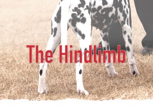
The Hindlimb
Click here to read the complete article
210 – January/February, 2022
 By Wendell Sammet
The hindquarters, or hindlimb assembly begins with the pelvis and the thigh bone (femur), which forms approximately 110 degree angle at its point of articulation with the ball and socket of the hip joint. At its distal end it articulates with the bones of the true leg, (tibia and fibula) at the knee (stifle joint). Distally, these two bones join to form the hock. The hind paw, or ankle, consists of seven tarsal bones (hock), five metatarsals (pastern), and the phalanges (the toes). Together, the many bones of the hindlimb complement the front assembly to create the dog’s running gear. Properly constructed and perfectly balanced, they efficiently produce and recycle energy with every step. A true marvel of engineering.
There are a lot of moving parts, so let’s break it down and examine each component.
The Pelvic Girdle
In terms of anatomical landmarks, the most prominent feature of the hindlimb assembly, the pelvis, is easily measured and evaluated. It serves multiple essential functions which include supporting and protecting the pelvic organs, forming a birth canal, and creating a solid base for the angulation of the hind limbs to produce the dog’s drive and propulsion. All things considered, a properly constructed pelvis is a vital element of soundness.
This large, bony structure is actually made up of two mirroring halves, a formation comparable to the fused pair of mandibles forming the lower jaw. Each half of the pelvis is comprised of three bones which fuse prior to birth, the ilium, the ischium and the pubis. When fused, their combined structure forms the deep socket of the hip joint, the acetabulum.
The Ilium
The ilium, the largest bony mass of the pelvis, is divided into two surfaces, three borders and three angles. Together they form the rounded wing at its cranial border of the pelvis, the iliac crest. This protrusion is a landmark for pelvic measurements. However, it varies in thickness depending on size of the breed, but in any case it can be easily observed and examined by touching the dog.
The bone of the ilium gradually thickens and widens to form a portion of the acetabulum, then tapers where it joins a narrow, more irregular shaped body, the ischium. Together they form most of the deeply recessed acetabulum, where the cylindrical head of the femur articulates, to form the ball and socket joint of the hip. The semicircular or half moon shape of the hip socket, called the lunate surface, is the weight bearing surface of the joint. The propulsive power of the hind limb is dependent on its strength and stability.
Pelvic Angle
Click here to read the complete article
210 – January/February, 2022

Short URL: https://caninechronicle.com/?p=221750
Comments are closed
