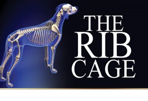
The Rib Cage
Click here to read the complete article
140 – March, 2022
 By Wendell Sammet
The rib cage is formed by thoracic vertebrae and the shaft of the rib is roughly oval with a distal end, somewhat enlarged, which is joined by cartilage. This is attached to nine small flat bones joined together by cartilaginous joints called “sternebrae.”
The first rib has, at its distal end, a projection called the “manubrium.” At the posterior is a triangular shaped projection called the “xiphoid process.”
The first 9 ribs are joined to the sternebrae. These are called the “true ribs.” The 9-12 ribs do not join the sternum but usually are attached to each other by cartilage, at the distal end of the sternal extremity. These 9-12 ribs are called the “false ribs.” The 13th is the true “floating rib.” When viewed in profile their formation represents the keel of a boat.
Rib Cage Thoracic Vertebrae
Click here to read the complete article
140 – March, 2022

Short URL: http://caninechronicle.com/?p=225462
Comments are closed


