Stem Cell Treatment for Spina Bifida Helps Dogs and Children at UC Davis
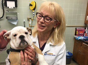
 A pair of English bulldog puppies born with spina bifida are the first patients to be successfully treated with a unique therapy—a combination of surgery and stem cells—developed at UC Davis by a team of veterinary and human medicine researchers and clinicians.
Dr. Dori Borjesson, professor of pathology, microbiology and immunology and director of the Veterinary Institute for Regenerative Cures (VIRC) at the UC Davis School of Veterinary Medicine, and Dr. Aijun Wang, assistant professor of surgery and co-director of the Surgical Bioengineering Laboratory (SBL) at the UC Davis School of Medicine, collaborate to identify diseases that affect both people and animals in order to pioneer unique regenerative medicine cures for these diseases. In the case of the bulldogs, placental stem cells were developed by the researchers, then characterized and combined with a tissue-engineered scaffolding to optimize treatment by UC Davis veterinary surgeons.
Because dogs born with spina bifida frequently have little control of their hind quarters, they also have little hope for a future. They are typically euthanized as puppies. Thanks to this groundbreaking research, there is now hope for these dogs, as well as a basis to inform human clinical trials for babies with the birth defect.
The surgical techniques, developed by Dr. Diana Farmer, professor and chief of surgery at the UC Davis School of Medicine, combined with the stem cell treatment developed by the research faculty, are progressions toward her goal of curing spina bifida. The disorder occurs when spinal tissue improperly fuses in utero, causing a range of cognitive, mobility, urinary, and bowel disabilities in about 1,500 to 2,000 children born in the U.S. each year. Accurate statistics on dogs born with spina bifida are not kept, as most are euthanized.
Dr. Farmer pioneered the use of surgery prior to birth to improve brain development in children with spina bifida. She later showed that prenatal surgery combined with placenta-derived mesenchymal stromal cells (PMSCs), held in place with a cellular scaffold, preserved the ability of lambs born with the disorder to walk without noticeable disability.
Dr. Beverly Sturges, professor of neurosurgery at the UC Davis veterinary hospital, wanted to find out if Farmer’s surgery-plus-stem-cell approach could give dogs closer-to-normal lives along with better chances of survival and adoption. At 10-weeks old, bulldog siblings Darla and Spanky were transported from the Southern California Bulldog Rescue to UC Davis, where they were the first dogs to receive the treatment.
After a few days of monitoring to see that they were healthy enough for the procedure, Darla underwent surgery on the third day of hospitalization, and Spanky on the fourth. In surgery, a myelomeningocele was identified at the L7 region of both dogs, consistent with the spina bifida identification on their MRIs. The dogs displayed dorsal positions of the lower extremities of their spinal cords, which were tethered. The sites were dissected and replaced in their spinal canals, with stem cells being surgically placed over the defects.
Prenatal diagnosis of spina bifida is not performed on dogs, which is why Darla and Spanky’s treatments occurred after birth, Dr. Sturges explained, as opposed to Dr. Farmer’s in utero approach. The disorder becomes apparent between one and two weeks of age, when puppies show hind-end weakness, poor muscle tone, incoordination, and abnormal use of their tails. At their 2-month recheck appointment, Darla and Spanky showed off their abilities to walk, run, and play.
“The initial results of the surgery are promising, as far as hind limb control,” said Dr. Sturges. “Both dogs seemed to have improved range of motion and control of their limbs.”
The dogs have since been adopted, and continue to do well at their new home in New Mexico.
Applying novel treatments to animals with naturally occurring diseases is making great strides toward helping to cure those diseases that occur in both humans and animals.
This “One Health” approach to medicine is best exemplified at universities like UC Davis, where physicians and veterinarians, along with their research counterparts, work side-by-side to develop cures and treatments to diseases and illnesses that affect humans and animals. The UC Davis team on this project included stem cell biologists and tissue engineers, along with the medical and veterinary clinicians – a group of forward-thinking minds that are not often found under the same roof.
The team hopes more dog breeders and rescues will send puppies with spina bifida to UC Davis for treatment and refinements that will help the researchers fix an additional hallmark of spina bifida – incontinence. While Darla and Spanky are very mobile and doing well on their feet, they still require diapers.
“Further analysis of their progress will determine if the surgery helped improve their incontinence conditions,” said Dr. Sturges.
Dr. Sturges hopes to start a canine clinical trial, which will further explore the possibilities of this treatment. With additional evaluation and U.S. Food and Drug Administration approval, Dr. Farmer also hopes to eventually test the therapy in human clinical trials. They hope the outcomes of their work help eradicate spina bifida in dogs and humans.
Funding for this project was provided by VIRC and SBL. Private donations to the veterinary school for stem cell research also contributed to this procedure. Drs. Farmer and Wang’s spina bifida research is supported by funding from the National Institutes of Health, the California Institute for Regenerative Medicine, Shriners Hospitals for Children, and the March of Dimes Foundation.
Short URL: http://caninechronicle.com/?p=131138
Comments are closed


