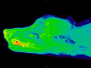
Advanced Imaging Puts UC Davis at Forefront of Cancer Treatment for Animals
 Cancer can be a devastating diagnosis for any pet owner. But now, thanks to new advanced imaging equipment known as the Mini Explorer II, UC Davis veterinarians can diagnose and treat disease earlier, with greater precision.
This imaging system—part positron emission tomography (PET) and part computed tomography (CT) scanner—is the first of its kind in the world and puts UC Davis at the forefront of cancer diagnostics and treatment. It will allow veterinarians to pioneer advancements in the coming All Species Imaging Center.
One of the most advantageous uses of PET in human medicine is to stage cancer, explained Dr. Erik Wisner, director of Imaging Services. While other forms of imaging (MRI, CT, radiographs, and ultrasound) are representative of imaging anatomy, nuclear medicine instead maps out physiologic or metabolic processes.
“Cancer cells behave differently than normal cells in terms of glucose metabolism,” Wisner said. “The most common radiopharmaceutical agent used in PET scanning behaves metabolically just like glucose, so it is taken up by cells. When one of those cells is cancerous, the agent becomes trapped in the cell, and it cannot be metabolized. A metastatic lesion in a lymph node might not be noticeable on a CT, but the agent trapped in the cell stands out on a PET scan.”
Through the Veterinary Center for Clinical Trials, Dr. Allison Zwingenberger is using the Mini Explorer II to conduct a carcinoma trial in dogs, examining a new imaging agent that may bind to mammary, colon, head, neck, pancreas, or prostate carcinomas, making the cancer easier to detect in the body.
Another advantage to the Mini Explorer II is the decreased time needed to conduct a scan. Typical PET scans can take up to 45 minutes to record the information needed for a diagnosis. Radiologists believe that the increased sensitivity of the new scanner may allow them to reduce this time to 5-10 minutes for some studies. This is possible because the number of PET detectors is much greater than a conventional scanner, allowing a larger volumetric acquisition. For example, the head, neck, and abdomen of a 30-pound dog can be scanned in two acquisitions instead of multiple readings required with a conventional scanner.
In another clinical trial, soft tissue surgeon Dr. Bill Culp is using the scanner to evaluate an alternative treatment for nasal tumors that eliminates the blood supply to the tumor, thus decreasing its size. This new treatment could help patients avoid lengthy radiation therapy and also lead to alternative treatments for humans.
In addition to cancer applications, the PET scanner is being used for orthopedic, neurology, and cardiology diagnosis, among others. Oral surgeon Dr. Boaz Arzi is conducting a trial on the scanner to assess its capabilities in determining the cause of vague jaw joint pain. These cases typically pose a diagnostic challenge because there are several disorders that can cause pain in that region. The precision of the PET/CT scan may help pinpoint the origin of the pain and determine a specific course of treatment.
The scanner (and its predecessor Mini Explorer I – currently at the Primate Center) was designed as a human model prototype by Department of Biomedical Engineering faculty member Simon Cherry with Ramsey Badawi, a physicist in the Department of Radiology at UC Davis Health. That human model, EXPLORER, the world’s first total-body PET scanner that can capture a 3D picture of the whole human body at once, is now up and running at UC Davis Health.
Short URL: https://caninechronicle.com/?p=171498
Comments are closed


