UC Davis Veterinary Orthopedic Surgeons Regrow Dog’s Leg Bone
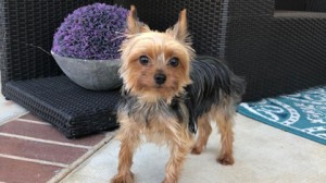
 A UC Davis veterinary patient is being described as a miracle by her owner. When Ethel, a 2-year-old Yorkshire terrier, was rescued by MaryAnn Lawson, the rambunctious pup was in a cast for a broken leg. Unfortunately, two previous surgeries failed to properly heal her broken right ulna and radius (equivalent to both bones in a human’s forearm). Lawson forged on and consulted with other veterinary orthopedic surgeons, all of whom recommended amputating the leg.
When Lawson asked the orthopedic specialists about other options, one mentioned a new procedure being used in veterinary medicine that involves regrowing bone with the use of bone morphogenetic protein (BMP), but felt that the procedure was not advanced enough to fix Ethel’s severe break. After leaving the consultation, Lawson started researching BMP and ultimately was referred to Dr. Amy Kapatkin with the Orthopedic Surgery Service at the UC Davis veterinary hospital. UC Davis has been utilizing BMP to regrow bones in dog jaws and legs since 2012.
Dr. Kapatkin agreed to take on the surgery but thought that there was only a one percent chance to save the leg. Although BMP grows bone effectively, the bone still needs to be stabilized with a bone plate to work. Ethel had 50 percent of her distal radius gone and her remaining bone was smaller than most bone plates made. Lawson agreed to allow Dr. Kapatkin to amputate once in surgery if it was determined that the use of BMP was not appropriate in Ethel’s case. But that turned out to not be necessary.
At UC Davis, Dr. Kapatkin has successfully implemented the regrowth strategy in nearly 25 dogs. All cases involved dogs like Ethel, who had nonunion fractures, meaning previous attempts to repair their bones failed to heal them.
“Nonunion after long-bone fracture repair in dogs represents a potentially devastating complication,” said Dr. Kapatkin. “When I first saw Ethel, I estimated that there was very little chance to save the leg – not impossible, though. And MaryAnn is such a dedicated pet owner that she insisted we try the regrowth procedure.”
While not all regrowth surgeries are the same, the basic premise of the procedure is to remove any dead bone and failed implants. A scaffold (called a compression resistant matrix [CRM]) saturated with BMP is placed into the bone defect to stimulate bone growth between the ends of the healthy native bone. Rigid stabilization (bone plate) of the normal bone on either end of the matrix with BMP has to be used. The use of CRM and BMP is specifically targeted for large bone defects that lack blood supply to the area after at least one failed surgery. Most cases, even those that had implants fail, can be managed with a second surgery and bone graft from the patient because the surrounding vascular environment is still healthy and only small defects are present.
“Like with almost all of the previous regrowth procedures, we had success with Ethel also,” said Dr. Kapatkin. “We are continuing to see exciting results with this procedure, and are optimistic that our use in orthopedics will have long-term positive results.”
Following surgery, Ethel was hospitalized at UC Davis for several months. Normally patients are able to be discharged in just a few days, but Lawson felt that Ethel’s surgery had the best chance for long-term success if she remained hospitalized under the watchful eye of UC Davis’ around-the-clock care team of veterinarians and technicians.
Normally, the bone plate used to stabilize the fracture remains in the patient forever. However, it was causing a problem with Ethel during her recovery. Due to the limited soft tissues and thin skin in her distal tiny limb, the plate started protruding from her leg. At a point in the recovery when the plate was no longer needed, Dr. Kapatkin removed it to allow Ethel’s tissue to completely heal around the healed bone.
At Ethel’s three-month recheck appointment, she was able to run on her new leg.
“I’m proud to be part of the process of what UC Davis is doing with this technology,” said Lawson. “This is important work that others can learn from so more dog owners can know this help is available. I’m so grateful to them and what they’ve done to help us.”
“Ethel has been such a blessing to us,” Lawson continued. “She’s our little miracle.”
Short URL: http://caninechronicle.com/?p=154731
Comments are closed


